 Introduction
Bone, or osseous tissue, is a specialized supportive connective tissue responsible for the support and attachment of soft tissues and organs (eg. muscle) (Junqueira et al., 2005). It also has a diverse range of functions, making it a very important tissue in the human body.
- Bone is responsible for the protection of soft tissues & organs, such as the heart and lungs in the thoracic cavity, or the brain encased by the cranium (Martini, 2006).
- Bone can function as a store of minerals (eg. calcium) and lipids from yellow bone marrow; this plays an important role in maintaining blood levels of ions and in osmoregulation of body fluids (Martini, 2006).
- Hematopoietic stem cells in bone marrow differentiate to produce erythrocytes, leukocytes, and platelets in the blood, to maintain appropriate levels for normal functioning (Martini, 2006).
- Many bones, especially the long bones of the limbs, provide leverage to change direction and magnitude of force applied by skeletal muscles, for movement of the body (Martini, 2006).

There are two types of bone present in long bones:
- compact (or cortical) bone
- spongy (or trabecular) bone
The bulbous end of the bone, or epiphysis, consists of spongy bone, with marrow filling the cavities between the trabeculae. The epiphysis is covered with articular cartilage, to reduce friction at the joints (Janqueira et al., 2005). There is also a thin region separating the epiphysis and the diaphysis, containing an epiphyseal cartilage, called the metaphysis (Martini, 2006).
Bone Histolgy
Bone is a dense, vascularized tissue composed of specialized bone cells in a calcified extracellular matrix containing fibers and ground substance (Janqueira et al., 2005).
Matrix:
- Predominant fiber = Type I collagen fibers
- ~ 1/3 of bone mass is collagen fibers
▸ very strong & can withstand tension
▸ flexible (can bend & twist)
▸ cannot withstand compression - Ground substance
- ~ 2/3 of bone mass is calcium phosphate, which reacts with calcium hydroxide to form crystals of hydroxyapatite. Other These crystals are hard, brittle, and rigid, thus they can withstand compression but are easy to shatter upon sudden impact or bending (Martini, 2006).
- Other calcium salts and inorganic ions such as Mg2+, and Na+ are also incorporated into the matrix.
- carbohydrates such as glycosaminoglycans
- proteins such as bone sialoprotein and osteocalcin
The mechanism of calcium phosphate deposition is not clearly understood, however it is known that the calcium salts are bound to proteoglycans and calcium-binding glycoproteins. Osteoblasts take-up and concentrate calcium in intracytoplasmic vesicles and release it into the matrix. Also, osteoblasts secrete alkaline phosphatase which aids calcification in some way (Janqueira et al.,2005).
Bone cells:
1. Osteoblasts
- responsible for bone growth.
- synthesize and secrete osteoid or pre-bone; the organic components of the matrix (Type I collagen fibers, proteoglycans, and glycoproteins) that have not yet been calcified.
- deposit inorganic components by increasing local calcium phosphate concentrations.
- located on the surface of bone tissue, and are closely apposed, resembling simple cuboidal epithelium (Junqueira et al., 2005).
- activity is stimulated by estrogens, androgens, growth hormone, and thyroxine.
2. Osteocytes
- derived from osteoblasts; those that have been completely surrounded with secreted bone matrix
- involved in the maintenance of the matrix (make and release proteins and organic matrix components) and are less active than osteoblasts.
- flat, almond-shaped, long-living cells
- located within lacunae; only one osteocyte is found per lacunae
- cytoplasmic processes lie within canaliculi and can make contact with other osteocytes via gap junctions (Junqueira et al., 2005).
3. Osteoprogenitor cells
- mesenchymal stem cells
- located in the cellular layer of the periosteum, in the endosteum that lines marrow cavities, and in the lining of blood vessel passageways through the matrix.
- divide to produce daughter cells which then differentiate into osteoblasts (Janqueira et al., 2005).
- important in repair of fractures (i.e. cracks in the bone) (Martini, 2006).
4. Osteoclasts
- large, multinucleate cells found in Howship's lacunae (Janqueira et al., 2005).
- responsible for resorption & recycling bone matrix by an erosion process termed osteolysis (Martini, 2006).
- secrete acids and proteolytic enzymes (eg. collagenase) to dissolve the matrix & release stored minerals (Janqueira et al., 2005).
- have projections and infoldings of the cell membrane forming a ruffled border which increases the surface area available for secretion of enzymes and resorption of bone (Janqueira et al., 2005).
- activity is stimulated by parathyroid hormone, and inhibited by calcitonin.

Fig.2. Morphological differences in bone cells (from http://www.lvh.org/assets/your_lvh/healthy_you/healthy_aging/bone_building.jpg)
- Compact Bone -
The collagen fibers and bone cells in mature, or secondary compact bone is organized into concentric lamellae which surround a central vascular canal containing blood vessels, nerves and loose connective tissue- referred to as a Haversian canal. There is also an inner circumferential lamellae around the marrow cavity, and an outer circumferential lamellae underneath the surrounding periosteum, each of which encircle the entire bone.
One Haversian canal and the surrounding lamellae is referred to as a Haversian system or and osteon. Other canals running perpendicular to the Haversian canals, called Volkmann canals, supply blood to osteons deeper in the tissue and to the tissues of the bone marrow cavity.
Between concentric lamellae are spaces within the calcified matrix called lacunae which contain one osteocyte each (Martini, 2006).
 Fig.3. Ground bone slide of compact bone (from http://www.feppd.org/ICB-Dent/campus/biomechanics_in_dentistry/ldv_data/img/bone_10.JPG)
 Canaliculi are narrow channels in the bone matrix that form a branching network through the lamellae and between lacunae and function in communication, nutrient exchange, and waste removal. Cytoplasmic extensions of osteocytes, called filipodia, accomodate these channels allowing a method of communication between neighbouring osteocytes, which are linked via gap junctions (Janqueira et al., 2005). The canaliculi also serve as a connection between lacunae and nourishing tissues, such as the marrow cavity, vessels of the Haversian canals, and the periosteum (Martini, 2006).
The periosteum is anchored to the surface of the osseous tissue by the penetration of collagen fibers, known as Sharpey's fibers. Fig.4. Structure of compact and spongy bone (from http://content.answers.com/main/content/wp/en-commons/thumb/f/f6/500px-Illu_compact_spongy_bone.jpg)
 Fig.4. Structure of compact and spongy bone (from http://content.answers.com/main/content/wp/en-commons/thumb/f/f6/500px-Illu_compact_spongy_bone.jpg)
- Spongy Bone -
Mature spongy bone also has concentric lamellae with lacunae and canaliculi housing osteocytes and their filipodia. However, the lamellae are not arranged into osteons.
Bone spicules form an open network of branching trabeculae, with bone marrow found within the spaces. Unlike compact bone, the bone matrix of the spicules are not vascularized - osteocytes must rely on diffusion of nutrients and wastes from the marrow and endostium lining along the penetrating canaliculi (Martini, 2006).
(from http://w3.ouhsc.edu/histology/Glass%20slides/69_02.jpg)
Fig.5. Spongy or trabecular bone
Osteogenesis
There are two different ways in which osseus tissue is formed:
- Intramembranous ossification (i.e. direct bone formation from mesenchymal stem cells)
- Endochondrial ossification (i.e. ossification of a preexisting cartilage template)
Both processes involved the production of primary, or woven bone, first; a primitive form of spongy bone. This type if osseous tissue is characterized by:
- large number of cells, a thick
- irregular network of collagen fibers
- irregular arrangement of lacunae (and hence, osteocytes).
- sutures of flat bones in the skull
- tooth sockets (gomphosis)
- some tendon insertions
Mature secondary, or lamellar bone is more organized and regular (see Fig.3.):
- lacunae are regularly arranged along lamellae
- collagen fibers are concentric and parallel within the lamellae
Mature bone is formed by either intramembranous or endochondrial ossification in different regions of the body (Janqueira et al., 2005).
1) Intramembranous ossification:
Occurs in the skull, clavical (collar bone), and part of the mandible (lower jaw) during embryonic development.
- Mesenchymal stem cells proliferate and aggregate in richly vascularized connective tissue (the primary ossification center), where they differentiate into osteoblasts
- Osteoblasts synthesize and secrete osteoid (or prebone) which form spicules that are calcified to form primitive or woven bone
- The spicules thicken & merge to produce a lattice network of spongy, or trabecular bone
- Spaces between the trabeculae contain loose, highly vascularized hematopoietic connective tissue that becomes primary bone marrow.
 Fig.6. Intramembranous bone formation (from http://www.engin.umich.edu/class/bme456/bonephys/osteogenesis1.gif)
2) Endochondrial Ossification:
Occurs in most long bones, vertebral column, ribs & pelvis during fetal development and continuing up until adolescence.
Endochondrial ossification requires a pre-existing hyaline cartilage models, that are ossified to become bones. Invading blood vessels are important for carrying the necessary cells to the center of ossification.
Involves two centers of ossification:
(i) Primary (diaphyseal) center of ossification
- Formation of a thin woven bony collar (periosteal collar) around the diaphysis by intramembranous ossification
- The perichondrium (i.e. the dense connective tissue layer surrounding cartilage; analogous to the periosteum) of the template becomes the periosteum
- Invasion of the diaphysis by blood vessels that carry osteoprogenitor cells from periosteum that mature into osteoblasts
- Osteoblasts secrete osteoid, and cartilage matrix begins to calcify
- Chondrocytes (i.e. cartilage cells) hypertrophy & die (because there is no diffusion of nutrients across the bone matrix)
- Osteoclasts form a primary marrow cavity, and incoming blood vessels carry in bone marrow cells
- Compact bone is formed (as described previously).
- Later, blood vessels infiltrate the epiphysis
- Chondrocytes of the epiphysis hypertrophy & die upon ossification
- Osteoblasts start building trabecular bone (Janqueira et al., 2005).
- Only a thin layer of the original hyaline cartilage remains on the surface of the epiphysis, forming the articular cartilage which prevents bone damage at the joints (Martini, 2006).
 Fig.7. Endochondrial bone formation (from http://www.cptc.ctc.edu/library/Bio%20118%20Lecture%20Notes%20Rev%200105_files/image048.jpg)
Bone Growth & Remodeling
Bone is a very dynamic tissue, continually undergoing reorganization and remodeling throughout an individuals life. The bone itself may appear unchanged, but the tissue is recycled by the regulated interactions of osteoblasts and osteocytes. In fact, approximately one-fifth of the adult skeleton is recycled each year. Bone may also change in size, shape, and general internal architecture. The largest changes in bone structure and size occur during the hormonal surge associated with puberty. Sex hormones (i.e. estrogens and androgens), growth hormone, and thyroxine all contribute to this dramatic change (Martini, 2006). Bone is capable of growing in both length and width- they typically lengthen early in development and widen after.
The epiphyseal growth plate is a narrow region of cartilage located in the metaphysis of long bones, which is responsible for the bone lengthening in young people, which can continue up until the age of 20. The proliferation of chondrocytes within this region leads to an increase in the length of the bone during endochondrial ossification. As they proliferate near the epiphysis, chondrocytes hypertrophy and die as their matrix becomes calcified by the action of osteoblasts near the diaphysis. As this occurs, the epiphyseal plate is being displaced (Janqueira et al., 2005). During puberty, osteoblasts are stimulated by sex hormones, growth hormone, and throxyine to produce bone faster than the cartilage can proliferate, leading to narrowing of the plate and epiphyseal closure (i.e. the epiphyseal cartilage will eventually disappear). When this occurs, the bones can no longer grow in length (Martini, 2006).
The epiphyseal growth plate contains 4 regions (Janqueira et al., 2005):
1) Reserve cartilage
- region closest to the epiphysis side; furthest from the zone of ossification
- resembles mature hyaline cartilage, without any morphological changes
- contains longitudinal columns of mitotically active cells (i.e. undergoing division) which grow in size
- also referred to as hyperplasia
- cells accumulate glycogen, and cytoplasm swells
- cells increase in size and increase the size of the lacunae as well.
- cartilaginous matrix becomes calcified by the deposition of hydroxyapatite
- chondrocytes die due the inhibition of nutrient diffusion across the calcified matrix
- forms the border between cartilage and the zone of bone deposition (i.e. ossification)

Fig.7. Cellular regions of the epiphyseal cartilage
(from http://www.med.mun.ca/anatomyts/msk/bone5.gif)

Fig.8. X-ray of a joint (arrows point to the epiphyseal growth plate)
from http://www.shoppingtrolley.net/images/epiphyseal-plate.gif)
Bones grow in width or diameter by the process of appositional growth.
Osteoprogenitor cells located in the inner cellular layer of the periosteum differentiate into osteoblasts, then secrete osteoid which is calcified to form bone. When the osteoblasts are completely surrounded by matrix, they differentiate in
As the bone grows outward, some of the older, deeper circumferential lamellae are remodeled, and replaced with osteons. It is also common for older osteons to be replaced by new ones by the deposition of lamellae by osteoblasts. When this occurs, the re

http://www.technion.ac.il/~mdcourse/274203/slides/Skeletal%20Tissues/19%20-%20Interstitial%20System%20-%20Lamellar%20bone.jpg
Pathology
A defect in any of the above mentioned components, and processes, characteristic of bone can lead to a variety of disorders. Here are a few:
1) Osteopenia: inadaquate ossification, leading to thinner weaker bones.
This is a normal part of the aging process. With age, osteoblast activity begins to decline, however, osteoclast activity remains the same; therefore, bones are undergoing erosion faster than new bone can be produced. Reduction in ossification begins between the ages of 30 and 40, and occurs more rapidly in women. Osteopenia is most obvious in the epiphysis of long bones, vertebrae, and jaws, leading to the weakening of the limbs, a reduction in height, and loss of teeth, respectively (Martini, 2006).
2) Osteoporosis: a reduction in bone mass to a degree that compromises normal function; essentially severe osteopenia.

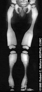
Rickets: a childhood disorder that reduces the amount of calcium salts in the skeleton, allowing bones to bend under the body's weight (e.g. "bow-legged").
Rickets is usually the result of a vitamen D3 deficiency. Thi
Flexibility of bones is due to the flexibility of the collagen fibers; the poor mineralization leads to decreased rigidity, and the bones' ability to withstand compression and tension. The femur bones thus bend laterally under the body's weight (Martini 2006). Rickets also causes the epiphyseal growth plate to become distorted, and bones become deformed (Janqueira et al., 2005). 
Fig.11. Anatomy of normal individual and one with rickets (from http://www.aurorahealthcare.org/healthgate/images/exh4511a_ma.jpg)
4) Osteitis fibrosa cystica: a condition in which increased osteoclast activity results in erosion of bone matrix and degeneration of fibers, due to excess parathyroid hormone secretion (Janqueira et al., 2005).
5) Osteopetrosis: a genetic disease in which osteoclasts lack a ruffled border; bone resorption is therefore defective.
It is not the direct effect of growth hormone that creates this effect, but its subsequent stimulation of somatomedins from the liver. During the years of growth, deficient growth hormone has effects on the epiphyseal cartilage that inhibits proper bone growth (Janqueira et al., 2005). Fig.13. A family with pituitary dwarfism (from http://trc.ucdavis.edu/biosci10v/bis10v/week4/4webimages/dwarf.gif)

7) Gigantism: immense bone growth resulting from an overproduction of growth hormone before puberty.
At this time, growth of the bone in length is uninhibited due to the presence of the epiphyseal growth plate (Martini, 2006). As for pituitary dwarfism, the condition is not due to the growth hormone directly, but by somatomedins from the liver (Janqueira et al., 2005).

Once the epiphyseal growth plate has closed, bone lengthening is inhibited, however, it can still increase in width by appositonal growth (Janqueira et al., 2005). The bones become very thick, leading to obvious deformaties of the face and hands (Martini, 2006). Fig.15. An individual with acromegaly (from http://www.sd-neurosurgeon.com/images/acromegaly%201.jpg)

9) Marfan's syndrome: a genetic condition linked to defective production of fibrillin, a connective tissue glycoprotein.
This deficiency of fibrillin leads to extreme height and long, slender limbs due to excessive epiphyseal cartilage formation, and cardiovascular problems due to the mutated structure of the connective tissues (Martini, 2006).

10) Scurvy: a condition involving weak, brittle bones as a result of a vitamen C deficiency in the diet. Vitamen C is an important cofactor for the enzymatic production of collagen, so other tissues, such as the skin, is affected as well (Martini, 2006).
It is also important to note that abnormal levels of particular hormones and vitamens, possibley due to another condition, have an affect on bone structure and function.
References:
Junqueira, L.C., and Jose Carnerio. Basic Histology: Text and atlas. 11th ed. USA: McGraw-Hill, 2005.
Martini, Frederic H.. Fundamentals of Anatomy & Physiology. 7th ed. San Francisco: Pearson Education Inc., 2006.